Case Studies & Research
Through advanced procedures, detailed case work, and ongoing learning, our team at Poulsbo Animal Clinic continually refines how we diagnose and treat complex conditions in dogs and cats. Discover informative examples of our surgical care and research presented here.
Surgical Case Studies: A Proven Record of Success
From orthopedic procedures to complex medical cases, these examples demonstrate how our team combines advanced techniques with carefully selected supplements to support meaningful outcomes.
Jaw Fracture Repair
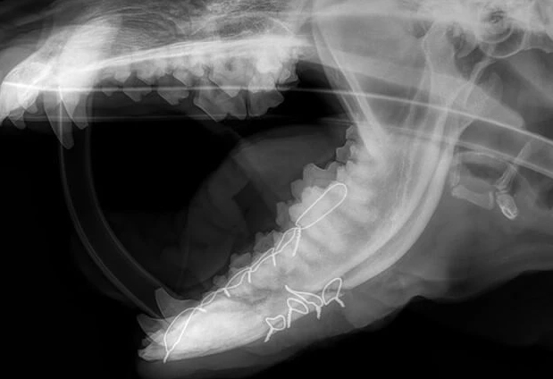
Booker is an 8-year-old German Shepherd that was kicked in the face by a horse on the family farm. He was diagnosed with a jaw fracture at the local emergency clinic and was sent to Poulsbo Animal Clinic for repair. He had an oblique fracture of the left mandible that involved a large butterfly fragment on the ventral surface and extended between the teeth into the mouth. Bone plates are typically not used on the jaw because the screws can damage healthy tooth roots. In addition, the repairs do not need to be as strong as long-bone fractures because the jaw does not support their weight as their legs do.
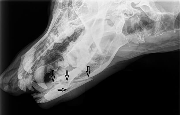
The butterfly fragment was pulled into apposition and affixed to the mandible with 4 wire loops called cerclage wires. In addition, an interdental wiring technique was used in which a wire is woven around the bases of the teeth and tightened. The protruding ends of the wire are splinted with a denture acrylic. This splint stayed in place for 8 weeks, after which the jaw had healed. Booker is back to running on the farm, hopefully steering clear of horse feet!
Sasha’s Excessive Tibial Plateau Slope
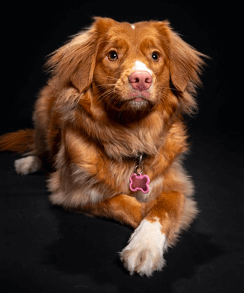
Sasha is a beautiful 2-year-old duck tolling retriever from Juneau, Alaska. She was diagnosed with a torn cruciate ligament in one of her knees, and the X-rays revealed excessive slope. Since it could not be repaired using the TPLO technique, her owner could not find a vet in Alaska who could do the repair. She flew from Juneau to have surgery in our clinic in Poulsbo.
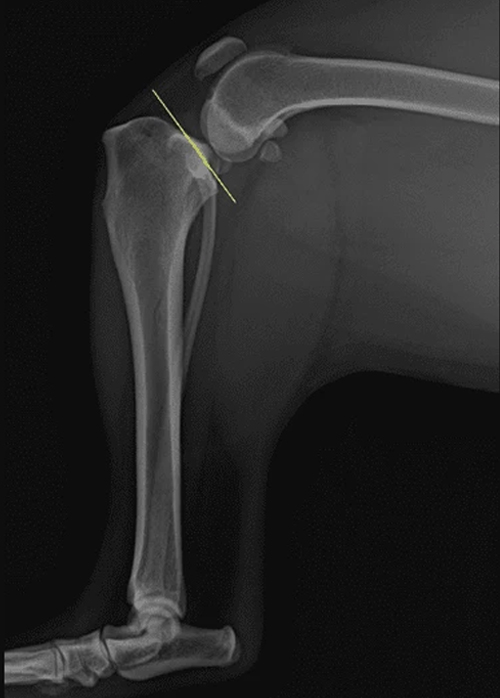
In the image above, the yellow line shows the excessive tibial plateau angle in Sasha’s left leg. This causes the femur to constantly want to slide off the back of the tibia. The cranial cruciate ligament stops the movement, but eventually, the excessive pressure on the cruciate ligament leads to its degeneration and subsequent rupture.
A traditional CBLO cut (image below – left) or TPLO cut (image below – right) would move the weight-bearing axis too far forward or too far rearward.
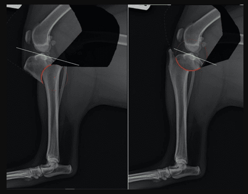
Now, refer to the image below. Using the double-cut technique, the red area in the photo to the left is removed prior to the forward rotation and fixation. The photo on the right shows the final pre-op plan for Sasha.
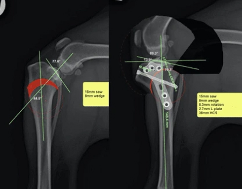
In the image below, Sasha’s post-op x-ray image shows the successful leveling of her tibial plateau using the double-cut CBLO technique at Poulsbo Animal Clinic by Dr. Adams.
CBLO Surgery: An Updated & Improved Version of the TPLO (Tibial Plateau Leveling Osteotomy)
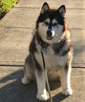
Duchess is a 7-year-old female malamute who is somewhat of a fixture on the Poulsbo waterfront, taking her owners, Ken and Amy, for walks multiple times per day. You might say she has achieved local star status when you see how many people know her by name. She started limping recently in her left rear leg and was diagnosed by Dr. Adams with a partially torn cranial (anterior) cruciate ligament in her left stifle (knee) that was confirmed on x-ray. Planning commenced for a CBLO (CORA-based leveling osteotomy).
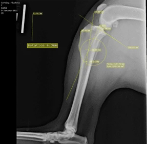
This is the X-ray image that was used for planning. The lines incorporate several landmarks on the bone that are used to calculate where to cut the bone based on the CORA (center of rotational axis), what size saw to use, how far to rotate the tibial plateau, and what size plate and screws to use.
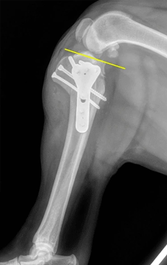
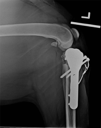
The post-op X-rays of Duchess reveal the placement of the plate and various screws. The wire that can be seen is a tension band that is not always needed, but in really large dogs, it adds an extra layer of stability as the bone is healing.
Three days post-op, Duchess is bearing weight very well. All of the dogs go home with special harnesses that allow us to support their weight and help them up until they are confident enough to do it on their own. They have 2 weeks of activity restriction, followed by 3 weeks of leash walking and physical therapy at home. Most are fully healed by 5 weeks and have no activity restrictions thereafter. The hardware usually stays in lifelong, but may be easily removed if indicated.
Little Bear’s Fractured Leg
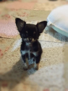
Little Bear is a brave little patient who came to us with a fractured front leg that had been splinted at an external provider. Unfortunately, issues with that splinting prevented poor Little Bear’s leg from healing properly, leaving a painful fracture remaining weeks longer than expected.
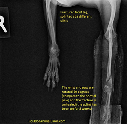
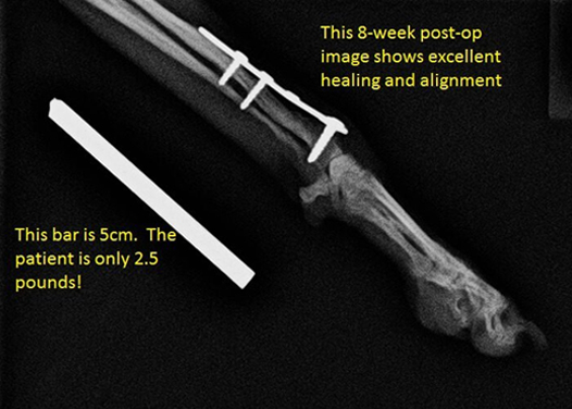
However, with some surgical help, we were able to give Little Bear bone plating positioned in such a way to support subsequent healing. We had to take particular care that our fix was as lightweight as possible to make sure we didn’t interfere with Little Bear’s recovery or day-to-day mobility.
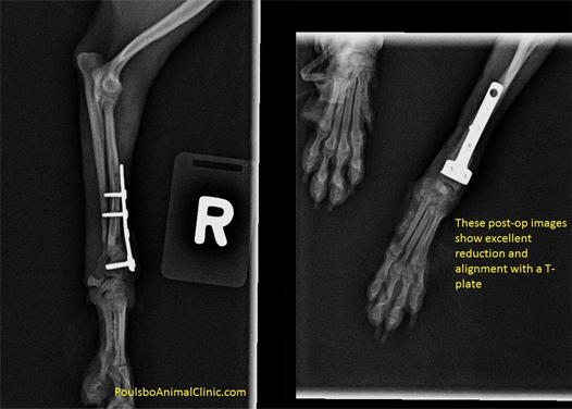
As you can see in the image below, after many weeks out, Little Bear’s prognosis was significantly improved.
Ear Surgery on Sundance the Guinea Pig
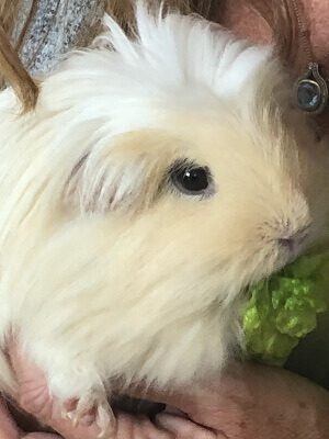
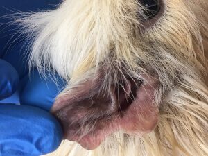
Sundance is a 1-year-old male guinea pig who presented to Poulsbo Animal Clinic with a chronic ear infection and red tissue growing out of the same ear. He had previously been treated with ear products and cleaners, but the presumed mass was making it difficult for the ear infection to completely heal. He was also scratching at his ear and was uncomfortable.
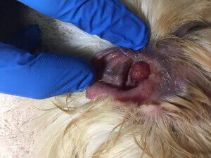
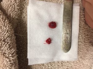
After further medical therapy was unsuccessful at improving the infection, odor, discharge, and his quality of life, his owners pursued surgery with us to remove the mass. We discussed that the red tissue could be a cancerous tumor, a benign tumor, or a nasopharyngeal polyp that may grow from the lining of the ear canal or eustachian tube. The traction surgical method was chosen as a first-line approach. If the tissue was cancerous or a polyp, the clients understood that it might regrow. To remove a cancerous tumor or a recurring nasopharyngeal polyp, a specialized surgery called a ventral bulla osteotomy may be required.
On the day of surgery, Sundance’s anesthesia and procedure were a success. The mass was removed via traction, bleeding was controlled, and the ear canal was flushed and treated. He recovered well from anesthesia and was back to his old self the next day at home. He never stopped eating!
He went home with additional product, and his ear was rechecked at our clinic two weeks later. No regrowth had occurred, and his ear looked and smelled like a normal guinea pig ear again. As of three months later, there’s been no regrowth. His family keeps a close eye on him and has enjoyed having their little buddy back and comfortable again!
Buddy's Double-Fracture Repair
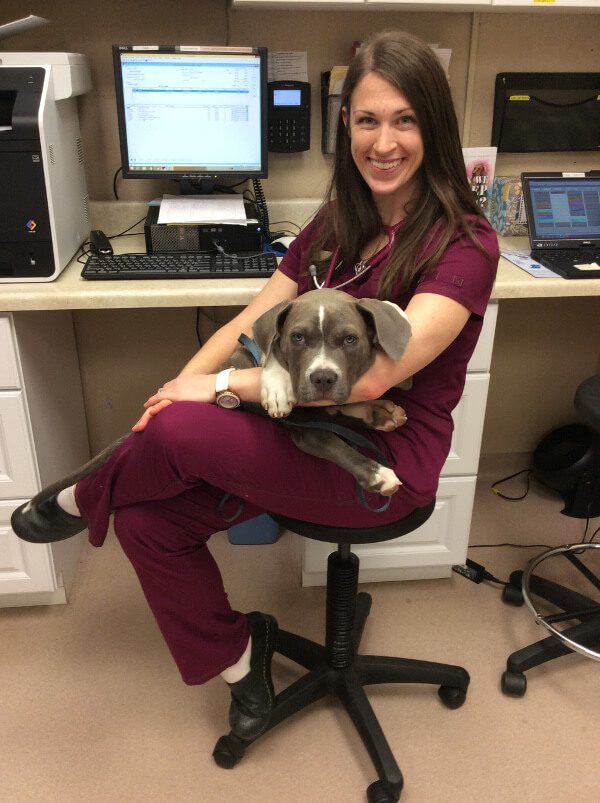
Buddy was seen at Poulsbo Animal Clinic as a 9-week-old puppy. He had been attacked by another dog and received life-threatening injuries. The owner first took him to a local emergency practice, where he was stabilized. They determined that his right femur and tibia were fractured during the attack, and they advised amputation (they even had a discussion about euthanasia).
Not wanting his young puppy to lose his leg, he found our website and saw that we perform orthopedic surgery. Buddy was examined, and we had a long discussion about his options. The owner decided that he would like us to attempt to repair the fractures and save the leg.
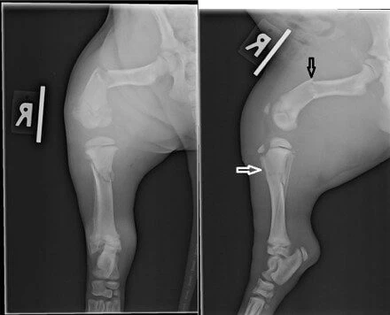
On the pre-op X-ray images, you can easily see the fractures.
The fractures were repaired using bone plates and intramedullary (IM) pins. The IM pins go down the center of the bones. They increase the strength of the repair by as much as 10-fold.
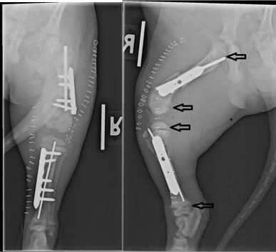
The black arrows on the second (lateral) image show the growth plates. The bones grow in length in these locations. Note how close the surgical implants are to the growth plates; the pin in the tibia even crosses the upper growth plate. The staple lines show that there were two separate surgical approaches: on the medial side to repair the tibia and the lateral side for the femur.
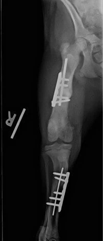
Buddy was kept quiet during the recovery, and the owner was very diligent about giving him his supplements. It is clear in his 8-week post-op X-ray images that he healed very well. Today, he is completely normal and uses his leg as if it were never injured!
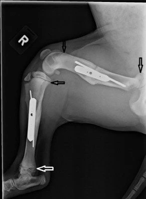
The arrows again point to the growth plates, but look how far they are from the implants now!
Research Papers: In-Depth Animal Studies
Our commitment to veterinary excellence extends beyond clinical care into research and scientific contributions. The following publications reflect a deeper exploration of animal health, behavior, diagnostic science, and the thoughtful use of supplements—offering insight into the principles that guide modern veterinary care. These studies highlight the importance of evidence-based care and the role of ongoing research in improving outcomes for animals.
Veterinary Clinic Pathology
This publication explores the foundational role of clinical pathology in veterinary products, focusing on how laboratory diagnostics—such as bloodwork, urinalysis, and cellular analysis—help veterinarians understand disease processes and guide treatment, including the appropriate use of supplements when indicated.
Scent Marking by Male Caribou: An Experimental Test of Rubbing Behavior
This research study examines scent-marking behavior in male caribou, exploring how factors like social status, environmental conditions, and physical characteristics influence rubbing behavior.